INVESTIGATIONS
There are various types of investigations that can assist your GP or Consultant Cardiologist to assess whether you have cardiovascular disease. Not all investigations are required to make the diagnosis of heart disease and this will be determined by your GP or Consultant.
Chest-pain Clinics
Patients suspected of having chest pain of cardiac origin are referred to chest-pain clinics by their GP. A full cardiac assessment and, if appropriate, an ETT (Exercise Tolerance Test or Treadmill Test) are taken at the clinic. Depending on the results, the patient is referred for the appropriate treatment or is discharged.
Blood Test
Measuring the fasting blood sugar for diabetes and cholesterol levels for hyperlipidaemia will help identify any undiagnosed risk factors. This is usually done by your G.P.
ECG – Electrocardiogram
 An ECG is a quick, painless and easy to perform test used to measure the electrical activity of the heart. Sticky pads are placed across your chest, wrists and ankles. These will be connected to leads and a machine will record the rate and rhythm of your heart. The ECG can provide valuable information about your heart. If it is not entirely normal, more tests may be needed.
Exercise Tolerance Test 
(Treadmill or Stress Test)
This test is sometimes called a stress test and it monitors your heart rate and blood pressure continuously while you are exercising on a treadmill. This test does not involve being admitted to hospital. You will be given an outpatient appointment to attend the hospital. You will need to wear clothing that you will be comfortable exercising in. Several sticky pads will be positioned on your chest and ECG leads will be connected to measure your heart rate and rhythm whilst you exercise on a treadmill. The recording of your blood pressure, heart rate and rhythm can allow your doctor to decide if further tests are needed.
Echocardiogram
Echocardiography is a helpful diagnostic imaging tool which uses ultrasound technology. This test uses sound waves to show the pumping action of the heart. It is also used to see whether if there is any damage to the heart muscle or valves. The equipment used is an advanced ultrasound  machine that can image your heart with the help of gel that is placed on the chest during the examination. This test does not require you to be admitted to hospital and you will be made an appointment to go to hospital as an outpatient.
Stress Echocardiogram
This is a similar examination to the echocardiogram and the same machine is used. In this test, the heart is made to beat faster and stronger using a special drug called “dobutamine”. This is injected into the arm whilst pictures of the heart are taken to see how the heart muscle responds to a faster and stronger heart beat. Some people feel slightly dizzy or nauseous during this test.
Myocardial Perfusion scan also known as a Thallium Scan or a Myoview scan
This test is used to observe the function of the heart muscle at rest and during exercise. This test does not involve admission to hospital. A set of pictures of your heart is taken when you are resting. The heart is also made to beat faster and harder either using physical exercise or using medication. At this point the second set of pictures of your heart is taken. A direct comparison of the heart muscle can then be made between rest and exercise. A Cardiac Perfusion MRI can also be used to assess the blood supply to different areas of the heart.
CT coronary angiogram
CT coronary angiogram involves taking pictures of the coronary arteries using x-rays. Narrowing in the coronary arteries can be identified early on with the use of CT imaging. Early detection can help to determine lifestyle adjustments and treatment options to avoid future heart disease.
CT uses X-ray exposure and women who are pregnant or potentially pregnant are advised not to have this scan.
Coronary Angiogram/Cardiac Catheterisation
Coronary angiography is carried out in a Cardiology Catheter Laboratory by a Cardiologist. A coronary angiogram offers a detailed record of the coronary arteries and their structure. You will need to be admitted to hospital in the morning and will be able to go home in the evening. The procedure takes approximately 30 minutes. This procedure is done under local anaesthetic, which means you will be awake. A needle is inserted into one of the blood vessels, either in the arm or in the groin. A small tube (catheter) is then advanced through the blood vessel and positioned at the opening of the heart arteries. A special dye is then injected into the arteries. This demonstrates any possible narrowing or blockages in the coronary arteries.
There are also many other investigations for a variety heart disease.
www.bhf.org.uk
These investigations will provide enough information for your doctor and your Consultant Cardiologist to decide:
 If your heart is healthy
If you need to start on medication
If you have narrowing or blockages in your coronary arteries
If you need an angioplasty to open the narrow areas in your coronary arteries
If you need to have open heart surgery
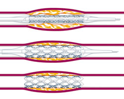
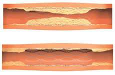
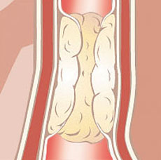 Narrowings or blockages in the coronary artery